
In Part 1, we learned the basic differences between bacteria and viruses so we had the background to understand the disease process of the microbes impacting dahlias. One of the ideas we discussed was that bacteria are ubiquitous. They are everywhere. We need to think about this because we often touch things thinking they are clean. The reality is we are picking up and depositing bacteria every time we touch a surface, or our plants. To prevent disease transmission from plant to plant, we need to be mindful of our hands as tools. Today, we are going to start with the simplest bacterial disease- Leafy gall.
A gall is any type of growth or lesion on a plant. In Leafy gall, the bacteria that causes this disease lives on the surface of the plant. It can be transmitted by gloves, snips, and other garden tools, but when it’s only on the surface of the plant, it doesn’t do much. Rhodococcus fascians is an epiphyte. It is a bacterium that can live on the surface of the plant without causing disease. That is because the stem and leaves create a barrier to the bacteria, like our skin. It is an opportunistic pathogen, meaning it can live on the surface of the plant without making galls, but if the plant stem gets torn, the bacteria can move into and across the stem. This is like you getting a cut and the bacteria on your skin moving into the deeper tissues.
This is where Leafy gall really gets going. The bacteria start to release signal chemicals inside the plant that disrupt the normal cells and alter the plant cell hormones. It causes the cells to grow in weird strands called fascicles (ribbons), which is where the scientific name comes from. Because the bacteria disrupt the hormones of the plant cells, the lesions and galls may present differently. This is why we see so many pictures with the question, “Is this gall?” Before we can appreciate all the ways Leafy gall presents, we need to understand how those precious eyes should form a nice healthy shoot.
Dahlias are part of a general plant group called dicots. These are essentially “not grass’s” and they grow with 2 initial leaves. Di means two. In a normal eye that develops into a sprout, the tip is a bit pointed with cells known as the meristem cells. These are the plant cells that are able to either become stem or leaves and through cell signaling, they will form layers to the stem.

Some layers become stem, others become leaves and in the above image, you may be able to pick out some of the two node branches that will become leaves from the stem. These tubers were forgotten in a box until fall and because they never got to sunlight, they did not trigger the hormone changes that cause chlorophyll to start to be produced and as such are white.
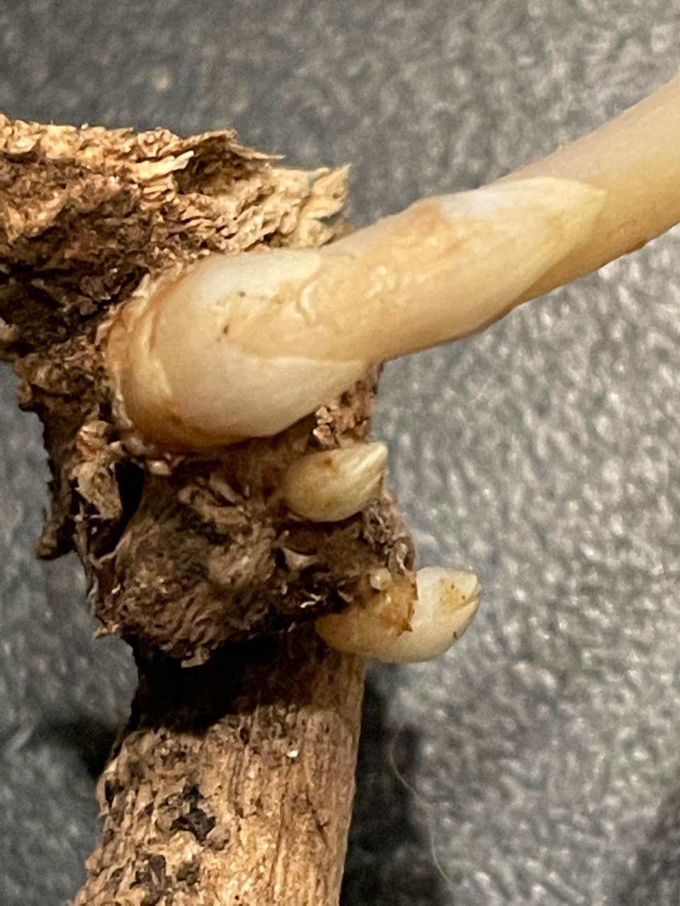
The eye and initial bud from the eye can be white or sometimes purple if the plant cells have hormones signaling pigment creation. A healthy tuber will have 1 to several eyes that are isolated from each other that form sprouts (picture to the left). The sprouts quickly take on a slightly pointed shape to press the soil away as they grow up towards the light. Without light, there is little signal for chlorophyll production and sprouts will be pale. This is important.
Leafy gall disrupts normal growth. It can start with a mass of “eye like tissue” all growing at the same time to create sprouts. This is what we associate as a gall. These bacteria that get into the plant don’t always make a visible gall. There are 3 typical presentations of Leafy gall: 1) tumors, 2) fascicle (ribbon) growth with fused stems, and 3) abnormal shoot development. In some plants, the hormones that would create that nice organized stem and leaf pattern gets disrupted and the sprouts seem rounded, curled, and or jagged. There are almost always more of these sprouts, and they seem short with folds of leaf buds tightly packed up the emerging stem. With the plant chemicals being messed with, it’s like the stems don’t know which direction to go.


Yet if other plant hormones are impacted, you might see dozens of tiny, fragile, hair like sprouts all white and densely clustered. Another off presentation is chlorophyll signaled in areas of the plant that are underground where the normal signal of sunlight is not there to cause chlorophyll production (see image below). Because the hormone signaling of the plant is affected, the way the plant presents its infection may look a little different depending on what state of growth the plant is in.


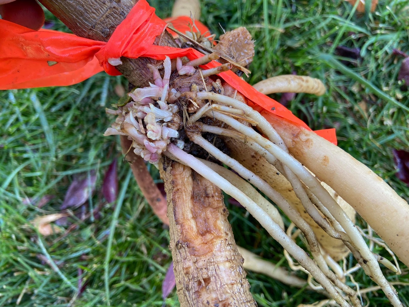
In this picture, you may notice thin tubers forming from the lesion site and the growing dense tissue of the lesion with stems that are going several different directions. The next picture is the same plant taken from a different angle. Note that the lesion is only on one side of the plant. Inspection of all sides of the plant are important and the bacteria can be found within this region, so all of this tissue, all sides of the plant are considered infected. The tubers on the unaffected side of the plant can not be saved.

There is some good and bad news with Leafy gall. This bacteria can be spread in your garden, but is probably the least transmissible (spreadable) in your garden. Studies have shown that when the bacteria get into the plant, the bacteria is only present in the few inches (15-25 cm estimate) of the gall. When the stem gets nicked, the bacteria enters that local area. There is no evidence to show these bacteria can move up the veins of the plants (in phloem or xylem). However, as an epiphyte the bacteria could be on any part of the plant, so if you have one area of the plant infected, it’s safe to consider the bacteria is on the surface of other areas of the plant. The general recommendation is if the plant does not develop a Leafy gall in 2 years, the plant likely does not have Leafy gall on its surface. Essentially, we are waiting to see if the plant gets a nick or cut that allows the bacteria on the surface into the plant to disrupt the plant’s hormones.
The bacteria living on any surface of the plant is why we see the disease present on any part of the plant. The bacteria has been located on seeds, leaves, stems, and the tuber. In the fall, a killing frost causes the stem and leaves to blacken and die. The bacteria on this part of the plant are done for the year. Most tubers are dug after a killing frost. If the weather improves, the plant attempts to regrow, so new eyes and stems start to grow from the tuber and buried remnants of the stalk. If we dig them in this time frame, we see the fall Leafy gall and these tubers should be destroyed. If the tubers were fully dormant when harvested in the fall and the bacteria are on the tuber, the process of dividing the tubers creates a cut on the surface that allows the bacteria into the space around the cells of the tuber. The following spring, when the plant starts to grow, the bacteria start to impact the tissues and we see the presentation of disease again. The tuber should be destroyed if it shows Leafy gall. We invest a lot into our gardens and dahlias; we don’t want to see all our plants diseased because something transfers the bacteria to our healthy plants.

Studies also show little support for insects transferring the bacteria from plant to plant in other plant species. This is likely due in part to the localized area often near the ground that the Leafy gall bacteria creates it’s lesions. One old previous study did find starved aphids transferred the bacteria to tobacco plants. While I have found mention of the study, I have not been able to find the actual report. There are two things to keep in mind about studies on this species of bacteria. Data specifically to dahlias is slim, but Leafy gall has been extensively studied in other plant species. Technology in science has made leaps and bounds in the last 10 years, so information from more recent studies holds more weight than studies that happened before we had the technologies for molecular studies. When an insect carries a disease, it is a vector. When vectors can carry disease, it’s a lot harder to control the spread and pollinators can rapidly spread disease. It’s a small miracle that Leafy gall is not spread widely by insects, but it can be spread by water spray.
If you plant new plants in your garden, they should be spaced far enough that rain, sprinklers, and wind do not cause one plant to touch its neighbor or splash water. Leafy gall transfer by water is well established. Studies have shown that infected plants in pots have bacteria in their water collecting saucers after the plants are watered. This is very important for all the gardeners who start tubers in pots or trays where water is able to spread from plant to plant by the underlying water catch tray. The studies found if you take the infected water from the catch tray and water a healthy plant, the water on the plant surface will infect the healthy plant. This means if you start your seedlings, starts, or tubers in 4 inch pots, and one is infected, even if it’s small enough not to touch its neighbors yet, the water that ran through can potentially contaminate all other pots that are in the same water catchment tray. Water splashing from one plant to its neighbor in the garden can transfer the bacteria. While water is a way to transfer Leafy gall, contamination of soil appears to be shorter lived.


The bacteria don’t appear to live in the soil as long as there is no residual plant material. Melodie Putnam, now retired, worked for Oregon State University’s Plant Clinic on this bacterium for years. She reported one study on how long bacteria can live in soil without a plant host was brief with dramatic decreases within 3 days of plant’s removal. She works with small herbaceous plants. A different study in pistachios (which are severely affected when diseases) discovered a dramatic decrease in bacteria in the soil over 2 months(2). However older studies reported persistence for 56 days. This confuses whether you should trust your soil after discovering an infected plan or not. Recall we have better technology to assess bacteria in soil, so the latest information gives us hope that soil is not long term contaminated.
As this bacterium is designed to survive the environment from year to year, the cautious gardener would not reuse soil and may remove that soil carefully. Leafy gall can be hosted in grasses (monocots) and broad leaf plants (dicots) there really is not a safe plant to plant in that place for a full year(1). Removing most of the plant material in the soil after digging your dahlias, coupled with the soil lying fallow without plants from fall to spring means there is a reduced chance of the bacteria being there in the spring. There really isn’t a way to make sure that all the little bits of roots and plant material is gone. If there is residual plant matter in the soil that the bacteria can live on, the bacteria might persist. It’s important to not compost infected plants as that compost could reintroduce the bacteria to the tubers you would plant in it.
Leafy gall lives on the surface of the plant waiting for an opportunity to get through the stem. Another study intentionally used snips on an infected plant and then stems from 10 healthy plants were nipped. When we use snips on a plant, this creates the break in the surface that the bacteria are looking for. Those snips used on other plants can carry the bacteria to new plants, so cleaning tools is an important part of preventing spread in your garden. The study found the 10 healthy plants did not get leafy galls, but a PCR test showed presence of the bacteria on those plants. A 10% bleach solution soak, Clorox wipes, or other antibacterial disinfectants should be considered for cleaning tools between use on different plants. Contact time with the disinfectants will impact their efficacy and if you only spend time to wipe your tool, you are at best decreasing the number of cells, but not eliminating cells. They should soak I. The disinfectant and depending on what you use, expect a minimum 5 minute soak or longer. It’s also important to note that the most effective disinfectants for dahlia viruses are bleach, Dawn soap, or Virkon S as reported in the March 2023 ADS/WSU Dahlia Virus Update talk. Viruses and bacteria are NOT the same type of life forms, and our cleaning products need to be specific for the bacteria or virus we are trying to target.
Near the base of the plant, using a rake or shovel that may nick the tuber is another way the bacteria can enter into the plant. Touching an infected plant bare handed or with gloves can transfer bacteria to you and then when you touch another plant, you spread the bacteria. Being mindful of how these bacteria may get transferred between plants will help break the chain of infection.
Leafy gall is possibly the most benign of all the dahlia diseases because it doesn’t stop the plant from growing. Dahlias from infected tubers may grow and bloom, or the stems may be weak and snap off. Yet removing these plants from the garden is important. The bacteria can be spread to many other plants in your garden including lilies and gladiolus. This bacterium is concerning for ornamental horticulture, but also in crops where it has significant issues in pistachios and beets. There is an attitude in some that this isn’t a big deal, but when we keep diseased stock, we spread the disease and different plants tolerate these bacteria differently. To be good stewards, and as painful as it may be, all diseased stock should be thrown out.

The takeaway message is any diseased plant risks your garden. The science on Leafy gall shows that water is the main way these bacteria spread where insects and soil are not as likely to spread the disease. Tools should be disinfected between plants and spacing plants should help minimize spread of the bacteria in the garden. With these disease transmission routes and good stewardship, suspected plants could be grown in isolation for observance. All sides of the plant should be inspected for infection and if a tumor, ribbon of growth, fused eyes and any abnormal growth is observed, the whole plant should be burned or thrown in the trash. As these bacteria that can live on all of the surfaces of the plant, but need to get through a wound on the surface of the plant to cause infection, all new plants should be isolated and observed for a minimum of two years. Leafy gall does not cause systemic infection of the plant like Crown gall does, but taking cuttings from infected plants means the bacteria can be on the surface of all the cuttings too. If you’ve found this interesting, then you will likely be fascinated with Part 3 on Crown gall. The infection process is more complex and severe, so while you may watch a suspected tuber with Leafy gall, when Crown gall is suspected the plant needs to be removed quickly.
Cheers and Happy Gardening
Special thanks to:
Thank you to Antonio Toledo, Lori Ratcliffe, Marie Willman, and Wendy Vreeken Banham for additional photos to provide this section with several photos of all the ways that Leafy gall can present.
Sources Cited in text:
(1)Plants that can and will host Leafy gall
Additional Reading:
Demystifying Rhodoccocus fasciens (2014)
Is it Crown gall or Leafy gall
Thank you for the info